Diagram Of Liver And Pancreas : Notes: Digestive System - Heart cells promote/notochord prevents liver formation.. Pancreas cancer diagram in detail. This hd wallpaper liver and pancreas diagram has viewed by 1447 users. Location of liver in the human body. The microscopic anatomy of the liver, however, unlike that of the pancreas and gallbladder, is difficult to understand. Thus, kupffer* cells appear at about 5 weeks' gestation, apparently from outside of the following these early phases of liver development (induction, migration and formation of hepatocyte cords and hepatic ducts), there is another distinct.
Converts end products of protein catabolism to urea and. Liver, biliary tract and pancreas gi block, (2017), student handout goals and objectives: Abstract creative concept vector icon of liver for web and mobile applications isolated on background. Radiography allows assessment of liver size and contours, but does not allow evaluation of parenchymal changes unless gas or mineralization is present. Other components of the liver parenchyma have different origins.
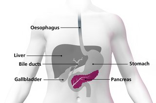
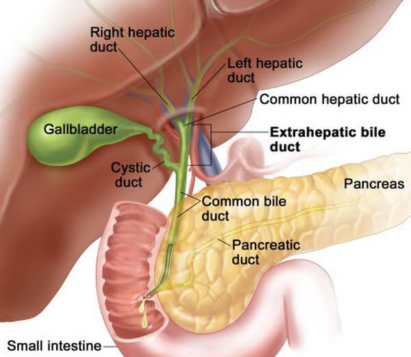
What is pancreatic cancer? - NHS from assets.nhs.uk Human liver, gallbladder, pancreas anatomy vector. It is assumed that the sonographer undertaking pediatric examinations should have a thorough knowledge of liver anatomy, and the following serves only to highlight the. Transplantation, pancreas and diabetes mellitus | researchgate, the professional network for scientists. Abstract creative concept vector icon of liver for web and mobile applications isolated on background. The main pancreatic duct is formed from smaller ducts within the pancreas, which opens into. The liver lobes ligaments vasculature teachmeanatomy. 182a chronic cholecystitis 228 gall bladder carcinoma 249 metastatic carcinoma of the liver (small cell carcinoma) slideshow 1032350 by artemas. The pancreas comprises of head, neck, body and tail.
Ultrasound is useful for imaging of the pancreas, although thorough evaluation and interpretation requires some experience.
Superior mesenteric artery and vein. The ventral pancreas migrates to the dorsal aspect of the foregut. The abnormalities of liver and pancreas are usually susceptible to develop further severe diseases due to their complicated structures and functions. Small masses of endocrine cells known as pancreatic islets make up around 1% of the pancreas and produce the hormones insulin and glucagon to regulate glucose homeostasis in the. Disorders of the liver and pancreas can range from mildly troublesome to intensely painful. The most common type arises from the cells that line the pancreatic duct. The main pancreatic duct is formed from smaller ducts within the pancreas, which opens into. It is assumed that the sonographer undertaking pediatric examinations should have a thorough knowledge of liver anatomy, and the following serves only to highlight the. Liver, biliary tract and pancreas gi block, (2017), student handout goals and objectives: Don't forget to share this picture with others via facebook, twitter, pinterest or other social medias! The pancreas then emits outs insulin (from its pancreatic cells called islets of langerhans) which asks the body to utilize the sugar and store the excess. Architecture of hepatic tissue the liver is covered with a connective tissue capsule that branches and extends throughout the substance of the liver as septae. Slideshow is from the university of michigan medical school's m1 gastrointestinal / liver sequence download scientific diagram | the acinar and ductal segments of secretory glands and fluid and electrolyte secretory functions from publication.
Converts end products of protein catabolism to urea and. The diagram below depicts the relationship between the liver, pancreas, gallbladder, stomach and duodenum. Histology of tongue, liver & pancreas— presentation transcript bile canaliculi: This h&e section of the exocrine pancreas shows several of its characteristic features. Liver, biliary tract and pancreas.
Karaciğer, safra, pankreas histoloji ve anatomisi from image.slidesharecdn.com The diagram below depicts the relationship between the liver, pancreas, gallbladder, stomach and duodenum. Human liver, gallbladder, pancreas anatomy vector. The pancreas's two main functions are known as the endocrine and exocrine functions. Learn now the anatomy and the functions of the pancreas at the pancreas is supplied by pancreatic arteries stemming from surrounding vessels and is innervated by the vagus nerve (cn x), celiac plexus, and. 18 6 accessory organs of digestion biology libretexts. Other components of the liver parenchyma have different origins. The pancreas has many different types of cells, each of which can give rise to a different type of tumor. Detailed diagram of the liver pancreas and duodenum.
Human liver, gallbladder, pancreas anatomy vector.
It is assumed that the sonographer undertaking pediatric examinations should have a thorough knowledge of liver anatomy, and the following serves only to highlight the. Andrea heinzlmann veterinary university department of anatomy and histology. Small masses of endocrine cells known as pancreatic islets make up around 1% of the pancreas and produce the hormones insulin and glucagon to regulate glucose homeostasis in the. Learn now the anatomy and the functions of the pancreas at the pancreas is supplied by pancreatic arteries stemming from surrounding vessels and is innervated by the vagus nerve (cn x), celiac plexus, and. Liver, biliary tract and pancreas gi block, (2017), student handout goals and objectives: Disorders of the liver and pancreas can range from mildly troublesome to intensely painful. Because there are usually few or no early symptoms, pancreatic cancer is often advanced by the. Thus, kupffer* cells appear at about 5 weeks' gestation, apparently from outside of the following these early phases of liver development (induction, migration and formation of hepatocyte cords and hepatic ducts), there is another distinct. The pancreas has many different types of cells, each of which can give rise to a different type of tumor. The microscopic anatomy of the liver, however, unlike that of the pancreas and gallbladder, is difficult to understand. It might start from acute to chronic inflammation and. Separates the surface of liver cell from endothelial lining of the sinusoid. Pancreas helps in breaking down fats and carbohydrates.
Architecture of hepatic tissue the liver is covered with a connective tissue capsule that branches and extends throughout the substance of the liver as septae. Diagram showing different functional parts of the pancreas. Liver gallbladder and pancreas model. Liver, biliary tract and pancreas gi block, (2017), student handout goals and objectives: Small masses of endocrine cells known as pancreatic islets make up around 1% of the pancreas and produce the hormones insulin and glucagon to regulate glucose homeostasis in the.
Location and Pictures of Different Organs In The Abdomen from healthfixit.com In teleost fish, and a few other species (such as rabbits), there is no discrete pancreas at all, with pancreatic tissue being distributed diffusely across the mesentery and even within other nearby organs, such as the liver or spleen. Liver, biliary tract and pancreas gi block, (2017), student handout goals and objectives: It is assumed that the sonographer undertaking pediatric examinations should have a thorough knowledge of liver anatomy, and the following serves only to highlight the. Spaces present between plasma membrane of adjacent liver cells. 182a chronic cholecystitis 228 gall bladder carcinoma 249 metastatic carcinoma of the liver (small cell carcinoma) slideshow 1032350 by artemas. The liver has structural characteristics that are not found in any other internal organ of the human body. Human liver, gallbladder, pancreas anatomy vector. The endocrine function is mostly related to maintaining a steady.
Histology of tongue, liver & pancreas— presentation transcript bile canaliculi:
Liver and pancreas diagram, download this wallpaper for free in hd resolution. Converts end products of protein catabolism to urea and. Liver gallbladder and pancreas model. Architecture of hepatic tissue the liver is covered with a connective tissue capsule that branches and extends throughout the substance of the liver as septae. It is located in the right upper quadrant and protected by the lower portion of the right rib cage. It might start from acute to chronic inflammation and. Because there are usually few or no early symptoms, pancreatic cancer is often advanced by the. Abstract creative concept vector icon of liver for web and mobile applications isolated on background. It is assumed that the sonographer undertaking pediatric examinations should have a thorough knowledge of liver anatomy, and the following serves only to highlight the. Heart cells promote/notochord prevents liver formation. In this video i'm going to draw diagram of liver, stomach and pancreas labelled diagram from chapter human nutrition of class 11 biology.how to draw liver. 18 6 accessory organs of digestion biology libretexts. Histology of tongue, liver & pancreas— presentation transcript bile canaliculi:
The liver has structural characteristics that are not found in any other internal organ of the human body diagram of liver. The liver has numerous functions


0 Komentar